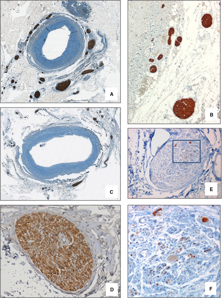
Figure 4.

Morphological findings of peri‐adventitial nerves. In patients in the DIAL group, a significant increase in nerve density in the internal area of the peri‐adventitial tissue (within the first 0.5 mm from the beginning of adventitia) was observed (A, NF stain—×2 and B, ×4) compared with the CTRL group (C, NF stain—×2). The efferent nerve fibers (D, tyrosine hydroxylase stain—×20) were more numerous than afferent fibers (E, calcitonin gene–related peptide stain—×20; F, Detail of area delimited by the rectangle in D, ×40). CTRL indicates control; DIAL, dialysis; NF, neurofilament protein.
