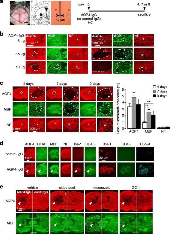
Fig. 3.

Mouse model of NMO for remyelination studies. a. Mice were infused with AQP4-IgG (or control IgG) containing human complement at day 0 using 40-μm tip diameter glass pipette and sacrificed at days 4, 7 or 9. Photographs show infusion (left), coordinates in striatum (middle) and glass pipette (right). b. Dose-determination study with indicated amounts of AQP4-IgG (each containing 30 % human complement) in a 3 μl total volume, showing AQP4, MBP and NF immunofluorescence at low (left) and high (right) magnifications. c. Time course of AQP4, MBP and NF immunofluorescence following intracerebral injection of 7.5 μg AQP4-IgG and 30 % human complement (left), with quantitative data (mean ± S.E., 6 mice, ** P < 0.01) (right). d. Brain sections from C were immunostained for indicated markers. e. Preliminary evaluation of potential remyelinating drugs (clobetasol 2 mg/kg/day, miconazole 10 mg/kg/day, GC-1 2 mg/kg/day). Mice were administered 7.5 μg AQP4-IgG or control-IgG and 30 % human complement at day 0 and treated with drugs (or vehicle) from days 1 to 9
