
Fig. 3.
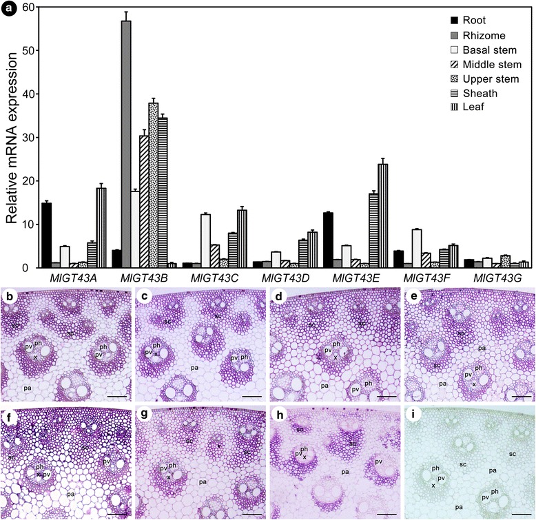
Expression patterns of MlGT43 genes. a Expression analysis of MlGT43 genes by qRT-PCR. Relative expression levels in seven tissues were normalized using MlACT11 as the reference gene. For each gene, the tissues with the lowest expression level are set to 1. Data are the means ± SE of three biological replicates. b In situ localization of MlGT43 genes in Miscanthus stem. Cross-sections of stems were hybridized with digoxigenin-labeled antisense MlGT43A (b), MlGT43B (c), MlGT43C (d), MlGT43D (e), MlGT43E (f), MlGT43F (g), MlGT43G (h), or sense (i) RNA probes, and the hybridization signals were detected with alkaline phosphatase-conjugated antibody and were shown as purple color. pv, pitted vessel; x, xylem; ph, phloem; pa, parenchyma; sc, sclerenchyma. Bar = 100 μm