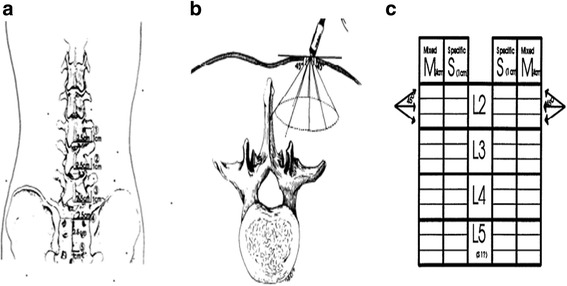
Fig. 1.

a With the patient prone, palpate 2.5 cm lateral and 1.0 cm cranial to the inferior edge of the spinous processes of L3, L4, and L5 and measure L2, L3, and L4 nerve roots, respectively. Mark a fourth location 2.5 cm lateral to the midline between the tips of the posterior superior iliac spines. Mark a fifth location 2.5 cm down to the midpoint and 1.0 cm lateral to the midline between the tips of the posterior superior iliac spines [20]. In this study, palpate 2.5 cm lateral and 1.0 cm cranial to the inferior edge of the spinous processes of L2 which was added to measure the L1 nerve root [21]. b Directions of needle insertion at each location. On the medial three insertions, the final l cm before contacting midline is scored “S” for specific. The remainder of these three insertions are scored “M” for medial. Note that the upper and lower medial insertions may not hit the spinous process before the needle hub touches skin, while the central medial insertion should do so if palpation was correct [20]. c The scoresheet: spontaneous activity is scored separately for insertions within the first 4 cm of insertion (placed in the M column on the scoresheet) and in the last l cm of insertion (placed in the S column of the scoresheet) [13]. In this study, L1 and S1 nerve roots were added to the scoresheet. PM scores were the summary of all plus at one nerve root level at one side
