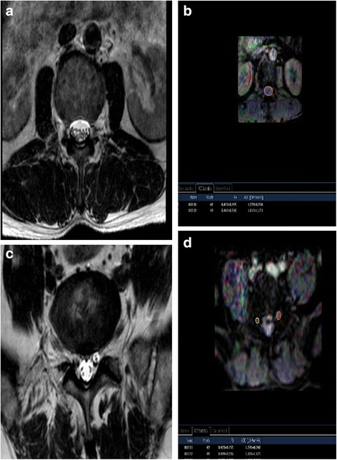
Fig. 3.

MRI T2W image of cauda equina (a) and FA mapping of DTI of cauda equina (b). ROIs were placed on the cauda equina on the zones equally as the disc, including superior 1/3, middle 1/3, and inferior 1/3 of the disc on FA mapping and FA values were calculated (b). The minimum values of three zones were taken as FA values of the cauda equina; MRI T2W image of bilateral nerve roots (c) and FA mapping of DTI of bilateral nerve roots (d). ROIs were placed on the “intraspinal,” “intraforaminal,” and “extraforaminal” zones of bilateral nerve roots on FA mapping and FA values were calculated (d). The minimum values of three zones were taken as FA values of the nerve roots. MRI magnetic resonance imaging, FA fractional anisotropy, DTI diffusion tensor imaging
