
Fig. 1.
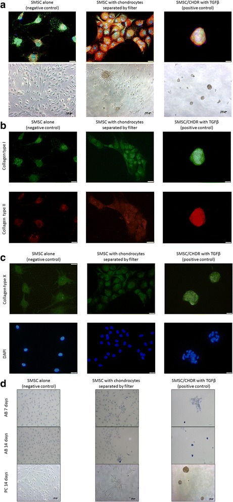
a Self-organization of SMSC after 7 days in monolayer culture. SMSC alone (left) stay separated, but in coculture with CHDR (middle) an aggregation of cells is visible, and addition of TGFβ (right) results in sphere formation. Upper row shows overlaying immunofluorescence staining (green: collagen type I, red: collagen type II, blue: DAPI; scale 20 μm), and lower row the phase-contrast microscopy. b Single staining for collagen type I (upper row) and type II (lower row) of the different groups. c Single staining for collagen type X and DAPI of the different groups. d Alcian blue (AB) staining of the different groups and time points. Upper row: AB 7 days (marker 50 μm), middle row: AB 14 days (marker 100 μm), lower row: phase-contrast (PC) microscopy 14 days (marker 100 μm). CHDR chondrocytes, SMSC synovial mesenchymal stem cells, TGFβ transforming growth factor beta (Color figure online)