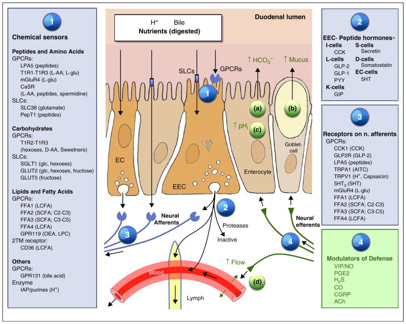
Figure 2.
Sensing, signaling pathways and duodenal epithelial defense. Sensing of luminal contents relies on G protein-coupled receptors (GPCR) and solute carriers (SLC; transporters) many of which are located at the apical brush border membrane (1) GPCR also known as seven-transmembrane receptors are cell surface receptors activated by a diverse range of inputs and ligands. Ligands bound to the extracellular face of the receptor activate intracellular G proteins, generating cascade-like downstream signaling pathways. SLCs exchange small solutes across plasma membranes or transfers solutes coupled to the transmembrane electrochemical gradient of ions such as Na+ or H+. Sensing is often linked to electrogenic activity that alters the transmembrane potential which subsequently enhances voltage-gated Ca2+ influx and subsequent Ca2+-induced stimulation of peptide hormone secretion. Binding of EEC sensors (1) in turn activates intracellular signaling pathways eventuating in the secretion of GI peptides or aromatic amines into the submucosal space (2). Released hormones act as paracrine mediators, can circulate systemically via blood flow or lymphatic flow, or are rapidly degraded. The release of GI peptides evokes local mucosal autocrine and paracrine mechanisms. Most of these signals are mediated through receptors in vagal, splanchnic and intrinsic afferent nerves (3). Mediators in efferent nerves evoke some or all factors in the duodenal mucosal defense system, including (a) stimulation of HCO3− secretion, (b) stimulation of mucus exocytosis resulting in a thicker mucus gel layer, (c) higher cellular buffering effort to protect against acid damage and (d) hyperemic response. For abbreviations, see Table 1. *Displayed to show classical EEC cell categories. Recent work suggests EEC may comprise a single cell type [3•,4•].

