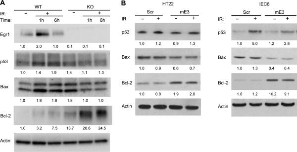
Figure 3. Knocking down Egr1 modifies the expression of apoptotic proteins.
(A) Egr1 WT and KO mouse pups were sham irradiated or cranially irradiated with 7 Gy. Total cellular lysate from hippocampal tissue was isolated 1 h and 6 h after irradiation. Cellular proteins were immunoblotted using antibodies against Egr1, p53, Bax, Bcl-2, and Actin (loading control). Densitometry values represent the ratio of the various proteins to Actin normalized to the WT 0Gy control. (B) Scr and knockdown HT22 and IEC6 cells were sham irradiated or irradiated with 4 or 6 Gy, respectively, and harvested after 6 h. Cellular proteins were immunoblotted using antibodies to p53, Bax, Bcl-2. Actin was used to evaluate protein loading. Densitometry values represent the ratio of the various proteins to Actin normalized to the Scr 0Gy control.

