. 2016 Feb 9;7(5):3414–3426. doi: 10.1039/c5sc04818d

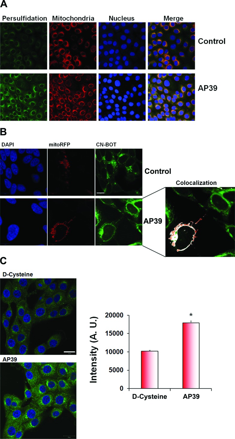
Fig. 4. Persulfidation co-localizes with mitochondria. (A) Co-localization studies of intracellular persulfidation. BAECs were transfected with RFP fused to the leader sequence of E1 alpha pyruvate dehydrogenase and incubated with mitochondria-targeted H2S donor, AP39. (B) SH-SY5Y neuroblastoma cells were transfected with CellLight® Mitochondria-RFP, BacMam 2.0 to visualize mitochondria and the cells were stained with improved tag-switch assay for persulfidation. Analysis with ImageJ (version 1.45R) revealed that significant co-localization was evident, which was visually represented as white pixels using the co-localization highlighter. Mitochondria-targeted H2S donor, AP39, lead to a strong increase in mitochondrial persulfidation. Scale bar 10 μm. (C) 100 nM AP39 is much more powerful inducer of persulfidation in the cells than 2 mM d-cysteine. Scale bar 20 μm.

Fig. 4