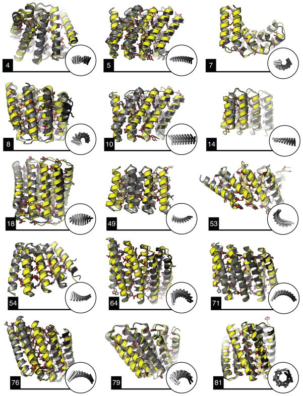
Figure 4. Crystal structures of fifteen designs are in close agreement with the design models.
Crystal structures are in yellow, and the design models in grey. Insets in circles show the overall shape of the repeat protein. The RMSD values across all backbone heavy atoms are: 1.50 Å (DHR4), 1.73 Å (DHR5), 1.30 Å (DHR7), 2.28 Å (DHR8), 1.79 Å (DHR10), 2.38 Å (DHR14), 1.21 Å (DHR18), 0.87 Å (DHR49), 1.33 Å (DHR53), 0.93 Å (DHR54), 1.54 Å (DHR64), 0.67 Å (DHR71), 1.73 Å (DHR76), 1.04 Å (DHR79), 0.65 Å (DHR81). Hydrophobic side chains in the crystal structures (in red) are largely captured by the designs (Extended Data Fig. 5).

