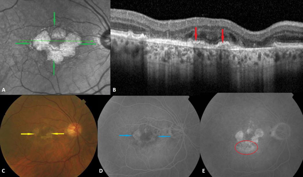
Fig. 1.
Combined geographic atrophy (GA) and choroidal neovascularization (CNV) in the right eye of a 75-year-old man. A. Multilobular GA is visible on infrared as small round or oval atrophic areas of increased reflectance (green arrows). B. Inactive CNV is shown on spectral domain optical coherence tomography as fibrovascular material below the retinal pigment epithelium (red arrows). C. The color photograph clearly shows the atrophic depigmented oval and round areas of GA (yellow arrows). D. GA is seen in the intermediate phase of the fluorescein angiogram as hyperfluorescent lobular lesions due to window defects (blue arrows). E. Occult CNV is characterized by an ill-defined area of irregular leakage and stippled hyperfluorescence in the late phase of the fluorescein angiogram (red circle).

