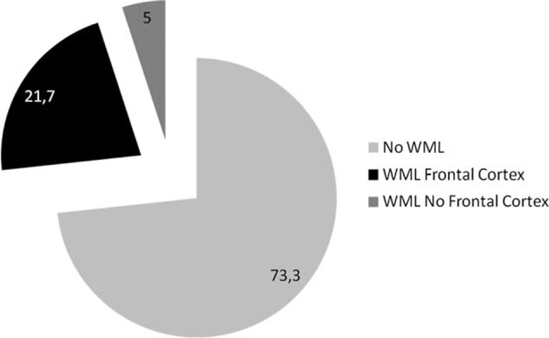
FIGURE 1.

Prevalence and topographic distribution of white matter lesions in the entire cohort of patients with fatty liver and controls without steatosis.

Prevalence and topographic distribution of white matter lesions in the entire cohort of patients with fatty liver and controls without steatosis.