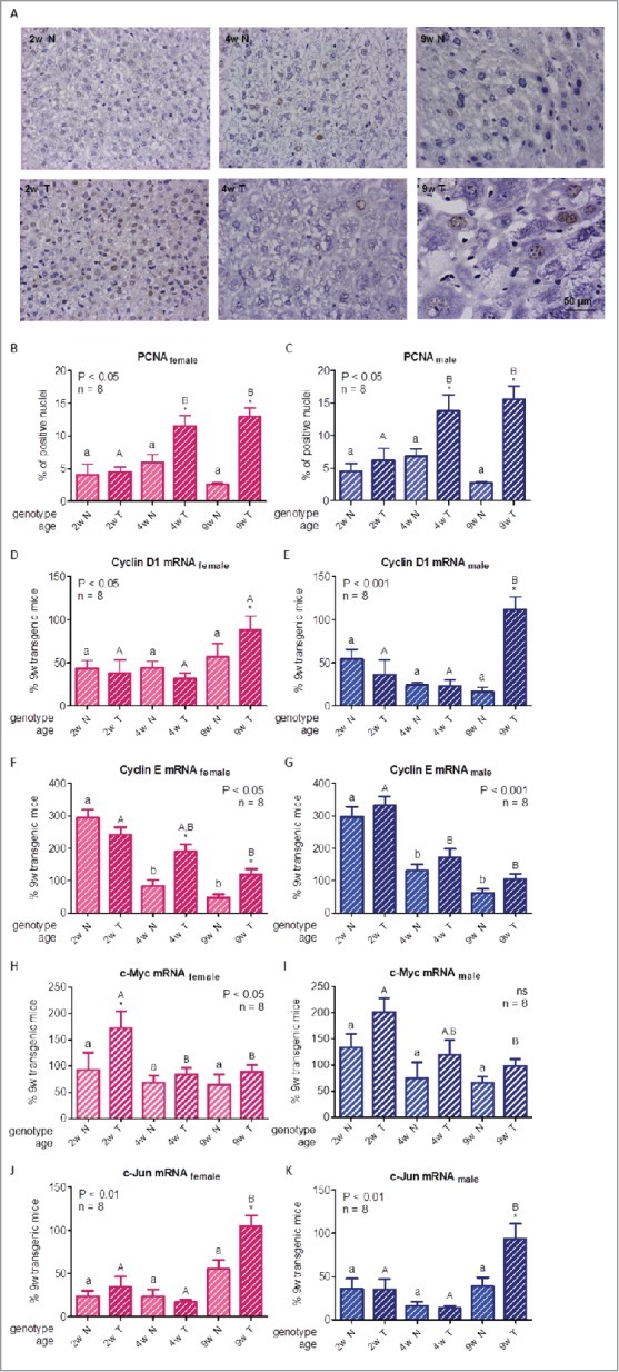
Figure 2.

For figure legend, see next page. Hepatocellular proliferation markers in growing GH-overexpressing transgenic mice and normal littermates. A: Representative photomicrographs of immunohistochemical staining of liver sections from normal and GH-transgenic male mice with anti-PCNA antibody; original magnification of 400x. B,C: PCNA immunostaining quantification in female and male mice liver respectively. D,E: Cyclin D1 mRNA hepatic expression in female and male mice. F,G: Cyclin E mRNA hepatic expression in female and male mice. H,I: c-Myc mRNA hepatic expression in female and male mice. J,K: c-Jun mRNA hepatic expression in female and male mice. Different cell cycle regulators and transcription factors involved in cellular proliferation were assessed in liver of GH-transgenic animals (T) and their non-transgenic littermates (N) for 2-week-old (2w), 4-week-old (4w) and 9-week-old (9w) mice. PCNA content was calculated as percentage of positive (brown stained) nuclei in 500 cells approximately. For simplification, only pictures from males are shown (A). To determine gene expression, mRNA was assessed by RT-qPCR from total RNA extracts. Values were related to cyclophilin A levels, referred to the average for 9-week-old transgenic female and male mice and expressed as percentage. Data are the mean ± SEM of the indicated n number of samples per group, each one representing a different animal. Different letters denote significant difference by age within genotype; small letters correspond to normal mice and capital letters to transgenic animals. Asterisks indicate significant difference between GH-overexpressing animals and their corresponding non-transgenic age controls.
