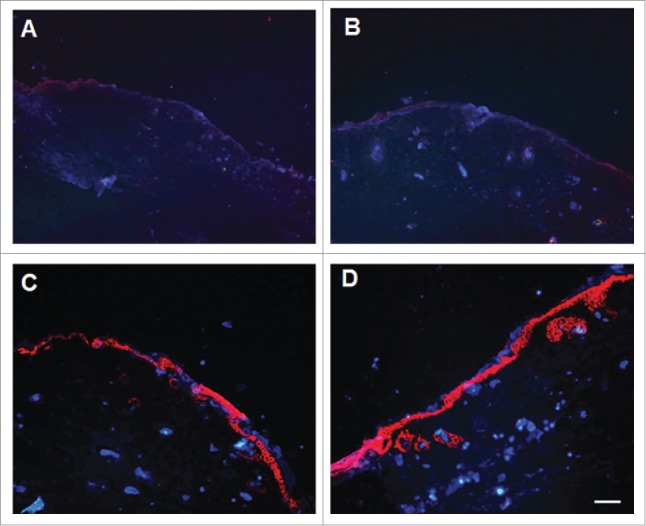
Figure 9.

Stratified epidermal layer on the wound surface formed by the implanted cells. A: Representative photograph of G-non group; B: Representative photograph of G-empty group; C: Representative photograph of G-CCND1 group; D: Representative photograph of G-positive group. Scale bar = 50 μm. CCND1, cyclin D1; G, group.
