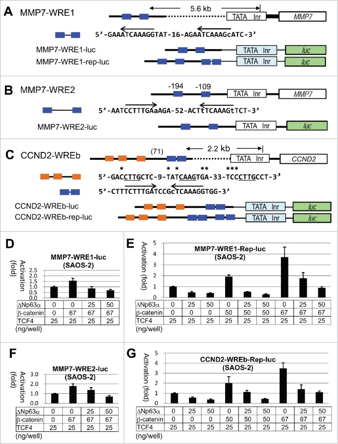
Figure 5.

Structural and functional analyses of WREs of MMP7 and CCND2.(A)-(C), Line drawings and nucleotide sequences for the WREs analyzed in this study. Endogenous promoter region containing TATA box (TATA) and initiator site (Inr), and the body of the gene are shown by white boxes. Bold lines mark the endogenous sequences, while the thin lines mark sequences in the plasmids. The light blue and green boxes signify the promoter and the luc gene in pGL3-promoter, respectively. Nucleotides matching the WRE consensus 32 in the positive and negative strands are indicated by arrows. Nucleotides deviated from the consensus are in lower case. (D)-(G), Results of the luc assays with SAOS-2 cells using the reporter plasmids shown in (A)-(C). Transfected plasmids and the DNA amounts are shown for each experiment.
