
Figure 5.
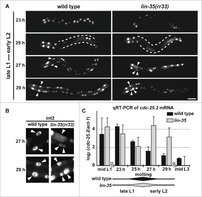
cdc-25.2 mRNA levels were negatively regulated by LIN-35. (A) GFP-marked intestinal nuclei in wild type and lin-35(rr33) mutants during early larval development. Hours (h) indicate the feeding period of synchronized L1 larvae in culture plates at 15°C. Dotted lines indicate regions of intestine in which GFP signals temporarily dispersed, and therefore intestinal nuclei were not clearly identified. Arrowheads indicate nuclei of int2 cells. Left, the anterior side. Scale bar, 25 μm. (B) Enlarged images of int2 nuclei. Excessive numbers of int2 nuclei were observed in 27 h- and 29 h-old lin-35(rr33) mutant early larvae (arrowheads). In contrast, the number of int2 nuclei was consistently 2 in wild-type larvae. (C) A time course of cdc-25.2 mRNA levels during early larval development in wild type and lin-35(rr33) mutants. The cdc-25.2 mRNA level at each time point in wild type and lin-35(rr33) mutants was measured 3 times by qRT-PCR, averaged, normalized to that of act-1 mRNA, and shown as the log2 value. Rhomboid shapes at the bottom indicate the duration of L1-to-L2 molting observed in wild type and lin-35(rr33) mutant larval samples.