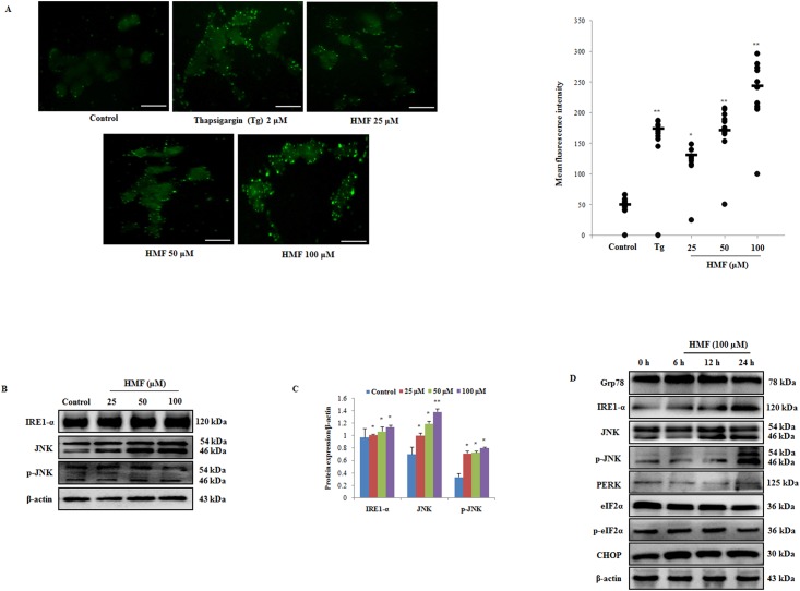
Fig 5. HMF-induced increase in cytosolic Ca2+and ER stress activation.
(A) Cells were loaded with fura-2 AM, and intracellular calcium release was observed after HMF treatment for 24 h. Thapsigargin (Tg) was used as a positive control. Positive rate of fura-2AM staining was analyzed by ImageJ software and presented as the mean ± SD (n = 10). (B) Western blot analysis of ER stress markers was performed using antibodies specific for IRE1-α, JNK, and p-JNK. β-actin was utilized as a loading control. (C) Densitometry analysis of respective proteins was carried out using ImageJ software, and results were normalized with β-actin in respective controls. (D) Determination of ER stress markers by HMF treatment in a time course manner was determined by western blotting. β-actin was utilized as a loading control. Data represent the mean ± SD (n = 3) from three independent experiments.*p<0.05; **p<0.01. Scale bars 0.1 mm.

