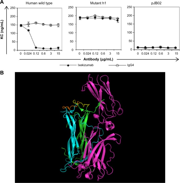
Figure 4.
Ixekizumab epitope.
Notes: (A) 4T1 cells were treated with a constant amount of human IL-17A wild type, mutant h1, or vector control supernatant in the presence of increasing amounts of ixekizumab (closed symbols) or isotope control antibody (open symbols). After 48 hours, KC in the supernatant was measured by ELISA. Results are shown as the mean of triplicate treatments ± SD and are representative of four independent experiments. (B) Structure of the IL-17A:IL-17RA complex (4hsa) with key amino acid residues in the epitope for ixekizumab highlighted. IL-17RA is colored in magenta, and the IL-17A dimer subunits are colored in cyan and green. The key epitope region (DGNVDYH) in IL-17A for ixekizumab is highlighted in yellow or brown in each subunit of the cytokine. This figure was generated using the PyMOL Molecular Graphics System (Version 1.7.0.3; Schrödinger, LLC).
Abbreviations: ELISA, enzyme-linked immunosorbent assay; IL, interleukin; SD, standard deviation; KC, keratinocyte chemoattractant.

