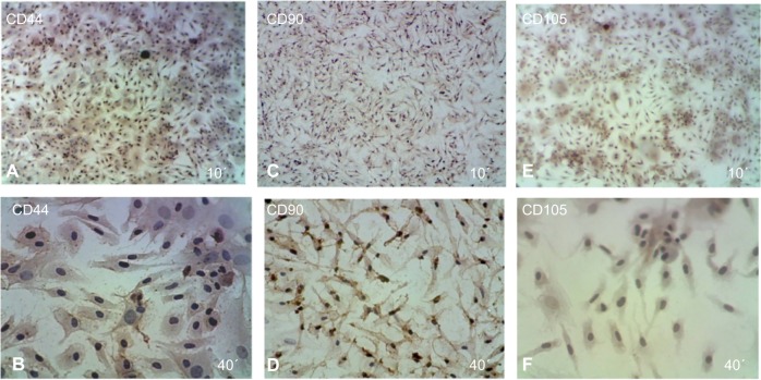
Figure 5.
Immunophenotypic analysis of MSCs at the first passage revealed under light microscope show that the MSCs were positive cells stained with brown color.
Notes: (A and B) CD44, (C and D) CD90, (E and F) CD105, note that all CDs are shown at 10× and 40×, respectively.
Abbreviation: MSCs, mesenchymal stem cells.

