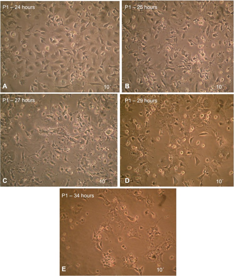
Figure 6.
MSCs after induced differentiation by BME, which showed the spherical shape of cells and their branched form toward the neural cells as revealed under inverted microscope.
Notes: All figures showed in 10×. The panels (A–E) were presented in 24–34 hours exposure times to differentiation media.
Abbreviations: MSCs, mesenchymal stem cells; BME, β-mercaptoethanol.

