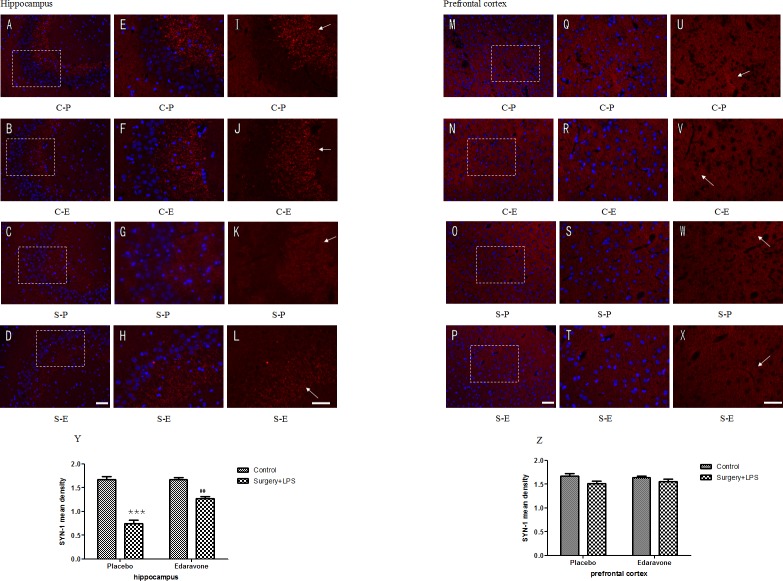
Fig 7.
Edaravone protected hippocampal and prefrontal cortical synaptic (red) integrity after surgery plus LPS administration (A-L) Representative images of SYN-labeled synapses in the hippocampi. (A-D) Synaptic protein and cell nuclei in the hippocampi on postoperative day 3 under a 200× magnification fluorescence microscope. (E-H) Synaptic protein and cell nuclei in the hippocampi on postoperative day 3 under a 400× magnification fluorescence microscope. (I-L) Synaptic protein in the hippocampi on postoperative day 3 under a 400× magnification fluorescence microscope. (M-P) Synaptic protein and cell nuclei in the prefrontal cortex on postoperative day 3 under a 200× magnification fluorescence microscope. (Q-T) Synaptic protein and cell nuclei in the prefrontal cortex on postoperative day 3 under a 400× magnification fluorescence microscope. (U-X) Synaptic protein in the prefrontal cortex on postoperative day 3 under a 400× magnification fluorescence microscope (Y) The density of hippocampal synaptic protein on postoperative day 3. (Z) The density of prefrontal cortical synaptic protein on postoperative day 3. Scale bars: A-D, 100 μm; E-L, 50 μm. ***P< 0.001 vs. C-P group; ##P <0.01 vs. S-P group. C-P, sham surgery plus placebo; C-E, sham surgery plus edaravone; S-P, surgery plus placebo; S-E, surgery plus edaravone.

