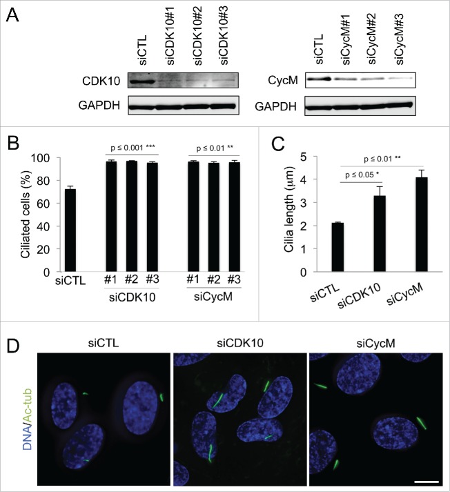
Figure 1.
CDK10/Cyclin M represses primary cilium assembly and growth (A-D) hTERT-RPE1 cells were transfected with control (siCTL), CDK10 or CycM siRNAs, subjected to serum starvation, and analyzed 72 hour post transfection. (A) CDK10 and CycM knockdown was assessed by western blot. (B) The percent of siCTL, si CDK10 and siCycM cells with primary cilia was determined by visualization using acetylated-tubulin staining (Figure S1A) and quantification. 500 cells were counted for each condition, in 3 independent experiments. (C) Measurement of cilia length (225 cilia/condition, in 3 independent experiments) using the LAS AF software on images randomly selected across the coverslips. (D) Representative immunofluorescence images of primary cilia (acetylated-tubulin staining, shown in green), DNA (DAPI staining, shown in blue). Scale bar: 10 μm.

