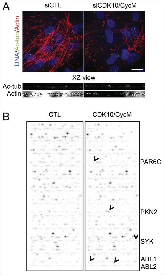
Figure 2.

CDK10/CycM maintains actin network architecture and phosphorylates actin dynamics regulators (A) Immunofluorescent visualization of primary cilia (acetylated-tubulin staining, shown in green) and F-actin (Rhodamine-Phalloidin staining, shown in red) without (siCTL) or with CDK10 or CycM silencing under the same conditions as Figure 1. DNA was stained with DAPI (shown in blue). Scale bar, 25 μm. The XZ view shows XZ optical projections of the cilia structure and actin structure (taken from a horizontal middle cross section) in a baso-apical manner. (B) In vitro kinase assay on protein arrays without kinase (CTL) or with recombinant purified CDK10/CycM. Positive hits (differential signals between both arrays) are indicated with black arrows and listed on the right.
