Figure 6.
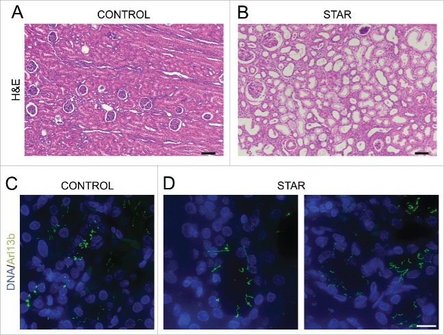
Renal tubules of a STAR patient are dilated and exhibit longer cilia (A, B) Histopathological analysis (H&E staining) of renal biopsies from a CONTROL (A) and a STAR patient (B). Scale bar, 100 μm. (C, D) Immunofluorescent visualization of primary cilia (Arl13b staining, shown in green) and DNA (DAPI staining, shown in blue) in renal tubules from a CONTROL (C) and a STAR patient (D). Scale bar, 15 μm.

