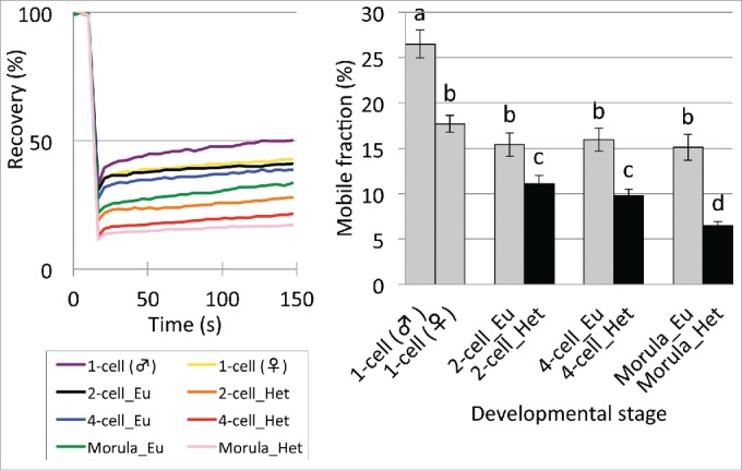
Figure 4.

Mobility of eGFP-H2B in euchromatin (Eu) and heterochromatin (Het) regions in preimplantation embryos. Embryos at the 1-, 2-, 4-, and morula stage were analyzed at 10–13, 28–32, 45–48, and 70–72 hpi, respectively. Heterochromatin regions were not analyzed in the 1-cell stage embryos because they were present only around the nucleolar precursor body at this stage. At the blastocyst stage, since the blastocoel made it difficult to distinguish heterochromatin from euchromatin, these regions were not analyzed. In the bar graph of the mobile fraction, different characters indicate statistical differences among developmental stages (P<0.05; by Student t-test) and the error bar indicates the standard error. More than 3 independent experiments were performed for each developmental stage and the data were accumulated. In total, more than 23 nuclei were examined at each developmental stage.
