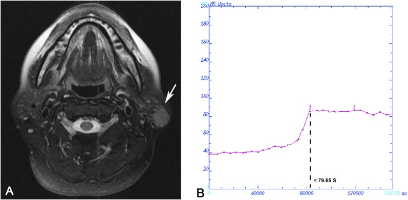
Figure 5.
Case 3: Tumour-like benign lymphoepithelial lesion in a 49-year-old female analysed by dynamic contrast-enhanced MRI. (a) Transverse T2 weighted spin time MRI showing a heterogeneously hyperintense solitary nodule in the left parotid gland (arrow). (b) Time–intensity curve shown as Type III, late increase type.

