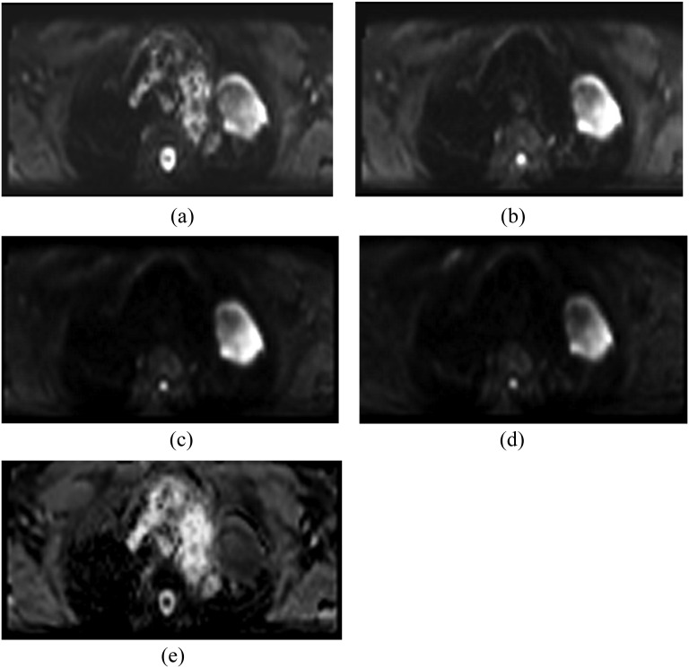
Figure 3.
Axial diffusion-weighted imaging (echoplanar imaging with ZOOMit) with b-value 0 (a), 250 (b), 500 (c) and 750 (d) of a 69-year-old male patient with lung cancer diagnosed with large-cell carcinoma of the left upper lobe. There is a marked area of hyperintensity in the left upper lobe characteristic of restricted diffusion. Artefacts are present, but the image quality is sufficient for tumour edge definition (d) and apparent diffusion coefficient measurement (e).

