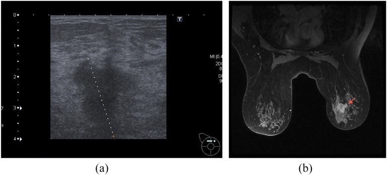
Figure 1.
(a) Ultrasound of the patient which was converted from breast-conserving surgery to mastectomy owing to lobular carcinoma in situ (LCIS); pre-operative ultrasound size 27 mm, MRI size 28 mm; histology showed 31 mm of invasive cancer and 57 mm of LCIS. (b) MRI of the above patient. Dotted line (a) and arrow (b) indicate ultrasound and MRI abnormality, respectively.

