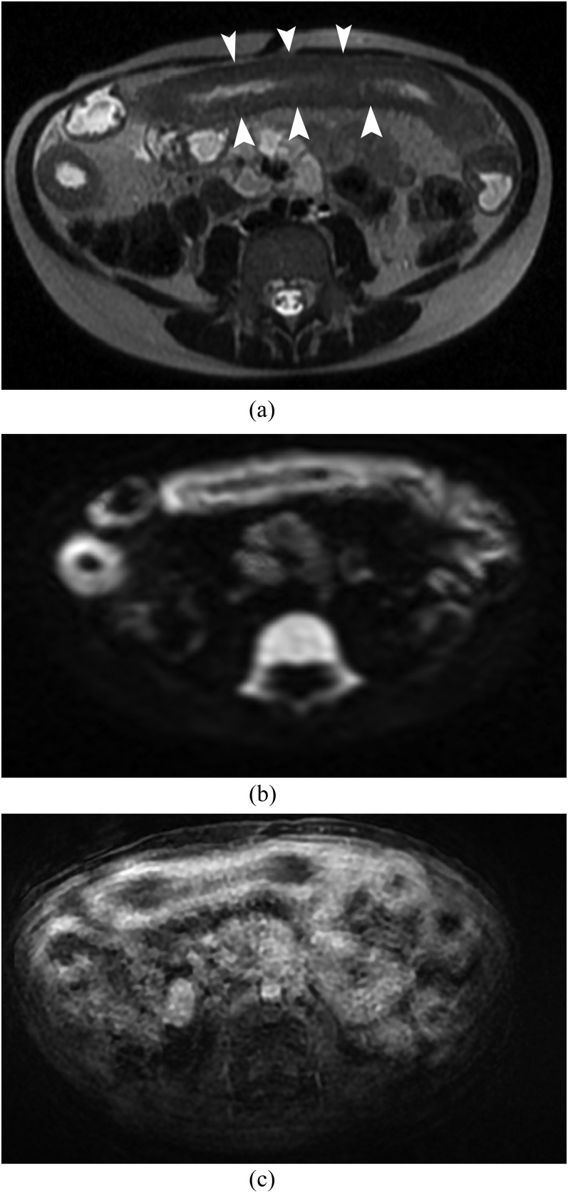
Figure 1.
MR enterography in 7-year-old female with Crohn’s disease. (a) Axial T2 single shot fast spin echo shows a long wall thickening (arrowheads) of the small bowel. (b) Axial diffusion-weighted imaging (b = 1000 s mm−2) shows a hyperintense signal of the small bowel with no artefact. (c) Axial T1 LAVA fat saturation after gadolinium enhancement shows a sequence with multiple artefacts. The enhancement is less well visualized as compared with DWI (b).

