Fig. 1.
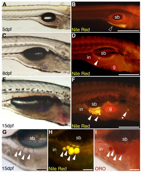
Fluorescent lipophilic dyes reveal lipid droplet accumulation in live zebrafish. Live zebrafish at 5 dpf (3.6 ± 0.1 mm SL), 8 dpf (4.3 ± 0.1 mm SL), or 15 dpf (5.3 ± 0.1 mm SL) were stained with the fluorescent lipophilic dye Nile Red, and imaged on a fluorescence stereomicroscope using a GFP longpass emission filter set. Bright field (A, C, E, G) and corresponding fluorescence images (B, D, F, H) are shown. Nile Red fluorescence emission maxima are shifted to shorter wavelengths when incorporated into neutral lipid, so neutral lipid depots in yolk (black arrowhead in B) and adipocytes (white arrowheads in D, F, H) appear yellow-orange. (A and B) The yolk is the major neutral lipid depot in 5 dpf larvae. (C and D) After yolk resorption, the first adipocyte neutral lipid droplets form in the right viscera by 8 dpf in association with the pancreas. (E and F) By 15 dpf, adipocyte lipid droplets have increased in number within the viscera, and also appear in other locations (asterisk in F). An individual 15 dpf zebrafish stained with Nile Red (G and H) then stained with Oil Red O (ORO; I) reveals colocalization of Nile Red and ORO staining in neutral lipid droplets of visceral adipocytes (white arrowheads). Swim bladder (sb), gall bladder (gb), and intestine (in) are indicated. Anterior is to the right, and dorsal to the top in all images. Scale bars: 400 mm (A and B), 300 mm (C–F), and 100 mm (G–I). This research was originally published in Flynn et al. (2009). © The American Society for Biochemistry and Molecular Biology.

