Fig. 5.
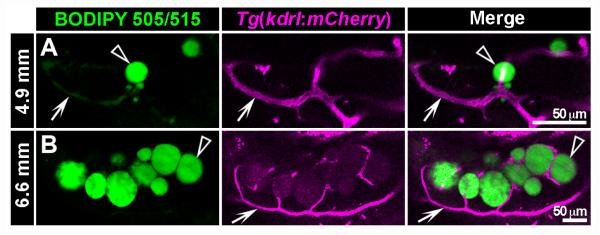
Confocal imaging of live zebrafish stained with BODIPY 505/515 fluorescent lipophilic dye reveals close interplay between lipid droplets and the vascular system in visceral adipose tissue. Tg(kdrl: mCherry) transgenic zebrafish express mCherry fluorescent protein specifically in endothelial cells of the vasculature (Jin et al., 2005). Live Tg(kdrl:mCherry) transgenic zebrafish were stained with BODIPY 505/515 according to protocol 1 (see Fig. 4) at 4.9 mm SL (rv14 dpf) and 6.6 mm SL (rv18 dpf). Stained zebrafish were imaged on a Zeiss LSM510 confocal microscope using filter settings for mCherry and BODIPY 505/515 (see Table I). Individual optical sections from a Z-stack are presented in this figure. At 4.9 mm, LDs (arrowheads) are small and loosely associated with vasculature (arrows, A). At 6.6 mm, LDs (arrowheads) are larger and interspersed with mCherry+ vasculature (arrows, B). Note: BODIPY 505/515 has weak fluorescence in the red channel (B), and also labels lipid in vasculature (A). Scale bars: 50 μm.

