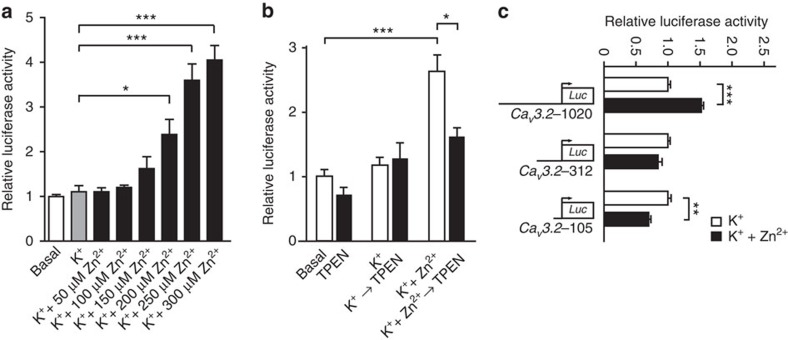
Figure 3. Increases in [Zn2+]i activate the CaV3.2 promoter.
(a) NG108-15 cells transfected with the 1,188-bp CaV3.2 promoter–luciferase reporter construct20 and stimulated with K++Zn2+ in the presence of increasing Zn2+ concentrations (0, 50, 100, 150, 200, 250 and 300 μM). The effect on the promoter activity was determined using a luciferase assay 4 h after stimulation (one-way analysis of variance (ANOVA): P<0.001; F(7,16)=25.64; Tukey's multiple comparisons test, *P≤0.05, ***P≤0.001; n=3). (b) Activity of the CaV3.2 promoter–luciferase reporter gene determined after stimulation of NG108-15 cells first with K+ or K++Zn2+ (50 mM/500 μM) solutions for 1 h and subsequently incubated in the presence or absence of TPEN (10 μM) for 1 h. Luciferase activity was measured 4 h after stimulation (one-way ANOVA: P<0.001; F(5,10)=24.63; Tukey's multiple comparisons test,*P≤0.05, ***P≤0.001; n≥3). (c) Luciferase activity of three CaV3.2 promoter deletion fragments20 after stimulation with K++Zn2+ (50 mM/200 μM). Only the CaV3.2-1020 deletion fragment showed significant activation of the CaV3.2 promoter after stimulation with K++Zn2+. The short CaV3.2-105 showed a significantly reduced activity after stimulation with K++Zn2+, likely due to the presence of Zn2+-inhibitory regions within this fragment (one-way ANOVA: P<0.001; F(5,12)=39.82; Tukey's multiple comparisons test, **P≤0.01,***P≤0.001; n=3).

