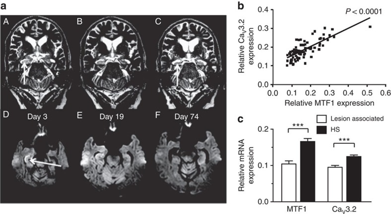
Figure 9. MTF1 and CaV3.2 expression levels co-segregate and are increased in hippocampal tissue of patients with HS.
(a) Epileptogenesis in a human individual without any previous neurological symptoms. The patient initially manifested clinically with SE (Supplementary Note 2). Coronal T2-weighted fast spin echo (A–C) and axial diffusion-weighted spin echo planar imaging (EPI) show the rapid development of a right-sided HS in the clinical course. Initially, there is hippocampal swelling (A: arrow) associated with cytotoxic oedema of the CA1 sector (D: arrow). Two weeks later, swelling and cytotoxic oedema are somewhat regredient but still present (B,E). Only 8 weeks later, cytotoxic oedema has disappeared (F) and hippocampal atrophy, that is, HS has manifested (C: arrow). MNI (Montreal Neurological Institute) coordinates as derived from the ‘standard brain template' correspond to 30, −14 and 20. (b) Regression analyses of CaV3.2 mRNA versus MTF1 mRNA expression in patients with HS. A strong positive correlation between the two variables is present even in the heterogeneous group of human HS hippocampi. (c) Quantitative determination of MTF1 and CaV3.2 mRNA. MTF1 and CaV3.2 are significantly abundant expressed in hippocampal tissue of TLE patients with HS versus hippocampi from patients with lesion-associated TLE, that is, in which seizures are explained by lesions such as low-grade neoplasms and/or focal dysplasia in the immediate vicinity or even including the hippocampal formation (HS: n=79; lesion associated: n=35; t-test: ***P≤0.001, with synaptophysin as reference gene).

