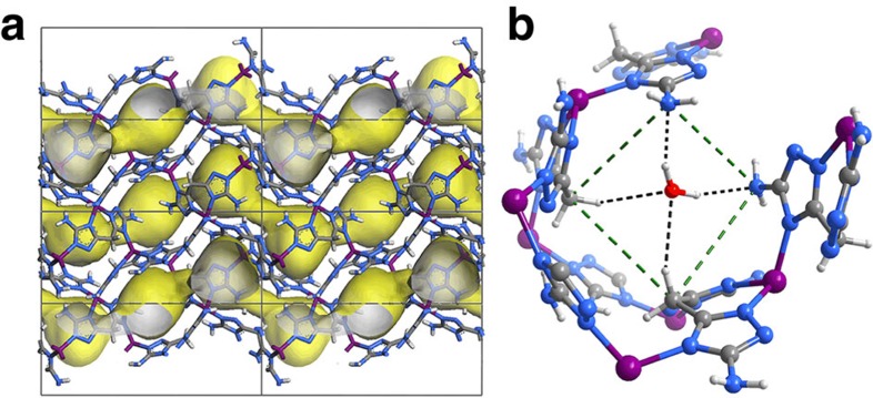
Figure 1. X-ray crystal structure of MAF-49·H2O.
(a) Framework (Zn purple, C dark grey, H light grey, N blue) and pore surface (yellow/grey curved surface) structures. Guest molecules are omitted for clarity. (b) Local environment and hydrogen-bonding interactions of the narrowest channel neck (highlighted by green dashed lines).

