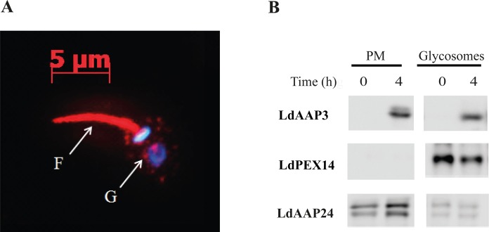
Fig 2. LdAAP3 localizes to both flagella surface and glycosome membranes.
A) Indirect immunofluorescence of LdAAP3. Axenic L. donovani promastigotes were stained using anti LdAAP3 IgG (red) and the DNA stain DAPI (blue). The latter stained the nucleus and kinetoplast. The two immunofluorescence images were merged. Immunofluorescence analysis was performed using the inverted cell observer Zeiss Axiovert 2. B) Subcellular distribution of LdAAP3. Axenic L. donovani promastigotes before and after 4 hours of arginine deprivation were extracted, non-soluble membranes were separated and subsequently subjected to sucrose gradient (20–60%) as described in Materials and Methods. Aliquots of plasma and glycosome membrane fractions before and after arginine deprivation were subjected to western blot analysis. Anti proline/alanine transporter (LdAAP24) and PEX14 were used as plasma and glycosome membrane markers, respectively. LdAAP3 molecular mass is 46 kDa. Note that length of exposure to peroxidase chemiluminescense was calibrated to starved cells. Bands in non-starved cells were visible after longer exposure (not shown).

