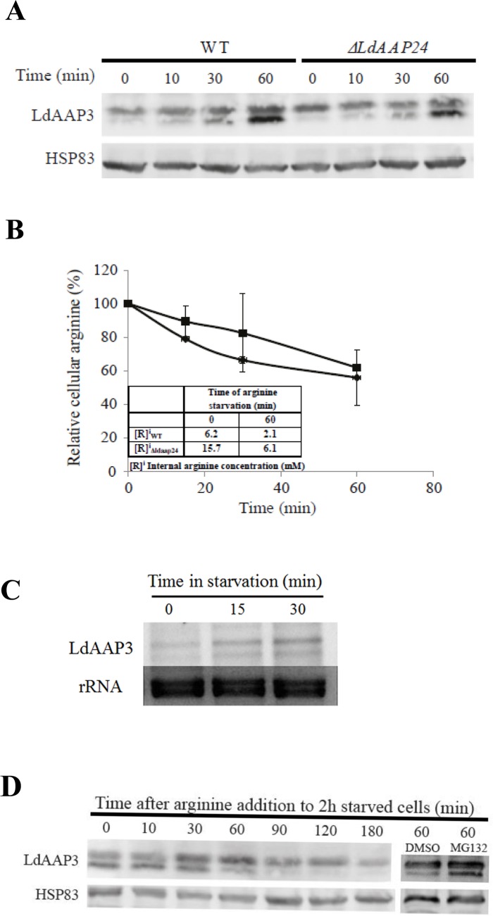
Fig 3. Time course of Arginine Deprivation Response (ADR).
A) Time-course of LdAAP3 protein abundance increase as function of arginine deprivation time in wild type (left four lanes, lower bands) and Δldaap24 (right four lanes, lower bands) L. donovani promastigotes. Mid-log phase parasites were suspended at a density of 0.5–1×10⁷ cells/ml in medium 199 without arginine and further incubated at 26°C. Aliquots were collected at 0, 10, 30 and 60 minutes, cellular proteins separated on 9% SDS-PAGE, transferred to nitrocellulose paper and subsequently probed with anti LdAAP3 antiserum. B) Rate of arginine pool depletion following arginine deprivation of L. donovani promastigotes wild type and Δldaap24. Aliquots of wild type (●) and Δldaap24 (■) collected in (A) were subjected to amino acid analyses as described in Materials and Methods. Values are mean ± S.D. (n = 3). Inset is a table that indicates intracellular arginine concentration values at zero and 60 minutes. C) Northern analysis of LdAAP3 mRNA abundance change during first 30 minutes of arginine deprivation. As a probe we used LdAAP3.2 (LinJ.31.0910). D) Arginine (0.45 mM) added to promastigotes after 2 hours of arginine deprivation induced rapid degradation of LdAAP3. Degradation was inhibited by the addition of 1 mM MG132 (Last two right lanes).

