
Figure 1. Domain arrangement and substrate specificity of drRecJ.
(A) Schematic of the domain arrangements of three DHH subfamilies. (B) Denaturing PAGE gel showing that drRecJ degrades different substrates, as shown at the top of the panel. 3′-Fluorescent labeled DNA or RNA (100 nM) were incubated with drRecJ (0, 5 and 20 nM) in the presence of 100 nM Mn2+ (see methods).

Figure 1—figure supplement 1. Sequence alignments, secondary structure, and functional residues of RecJ and CDC45.

Names of species are dra, Deinococcus radiodurans; ttj, Thermus thermophilus; eco, Escherichia Coli; Homo, Homo sapiens; Drosophila, Drosophila melanogaster. Structural elements of RecJ are shown in distinct colors. Predicted secondary structure of human Cdc45 is shown at bottom (grey). Conserved motifs (I-VII) are labeled. Conserved key residues are highlighted in distinct colors.
Figure 1—figure supplement 2. Metal preference of drRecJ.
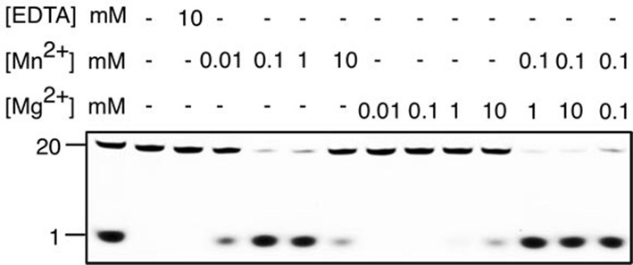
3′-fluorescent labeled 20 nt ssDNA (KY08, 100 nM) was incubated with drRecJ (10 nM) in the presence of Mn2+ and Mg2+. For the metal competition assays, drRecJ was pre-incubated with 0.1 mM Mn2+ before addition of Mg2+.