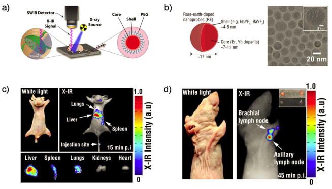
Figure 1.
(a) Schematic of REs showing the lanthanide-doped core surrounded by an undoped shell. (b) TEM images of REs reveal spherical morphology. (c) Nanoprobe clearance visualized in mice 15 min post injection using X-IR imaging. (d) X-IR imaging of axillary and brachial lymph nodes. Reproduced with permission from [65]. Copyright 2015 American Chemical Society.

