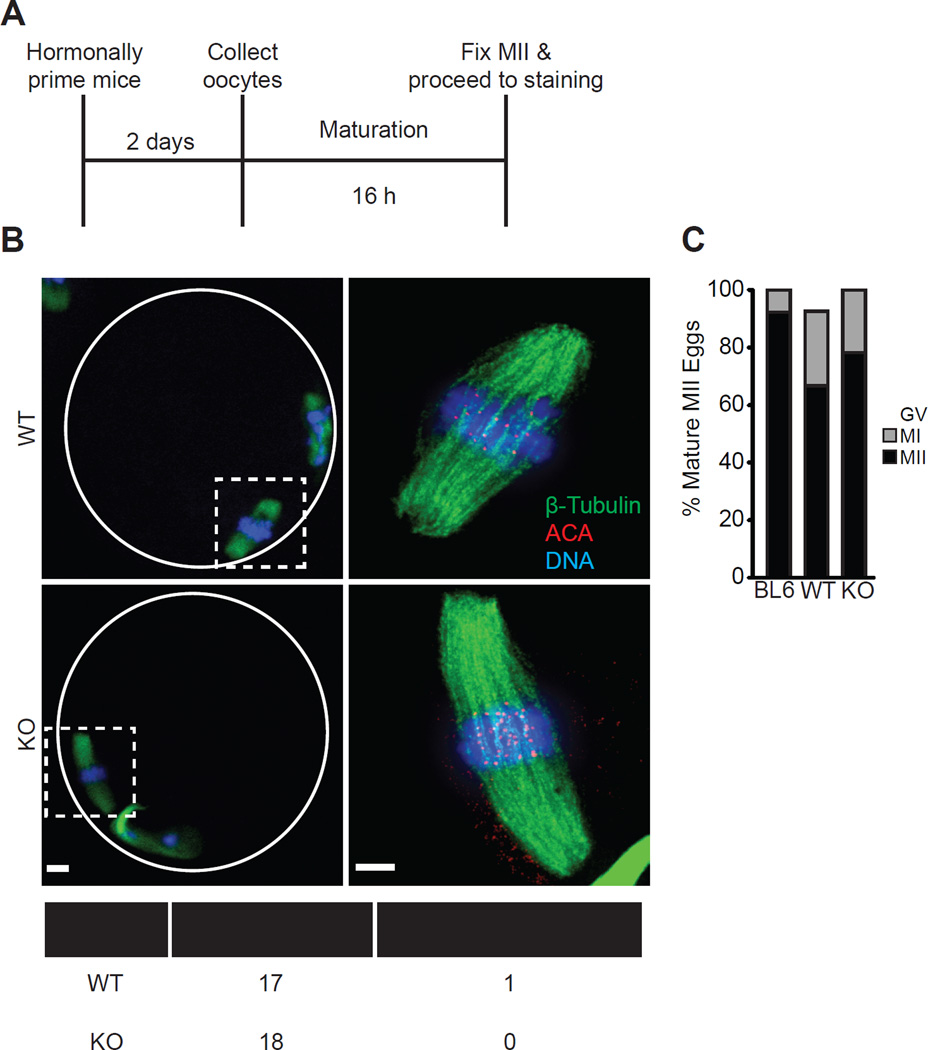
Figure 3. CENP-A nucleosomes assembled early in prophase I support normal meiotic centromere function.
(A–C) Oocytes were collected from 12 month old WT and KO females, or young C57BL/6J controls, matured in vitro to MII, and stained for β-tubulin, ACA to label centromeres, and DNA (schematic, A). Representative images (B) show MII eggs at 20X magnification (scale bar 10 µm), white circle represents zona pellucida; dashed squares are regions imaged at 100x (scale bar 5 µm). The percent in each group that remained arrested with an intact germinal vesicle (GV), arrested at metaphase I, or progressed normally to MII was quantified (C, n=27 oocytes from 3 WT mice, 23 oocytes from 3 KO mice, or 26 oocytes from 2 C57BL/6J (BL6) mice). The table summarizes how many MII eggs from WT and KO females had chromosomes aligned at metaphase.

