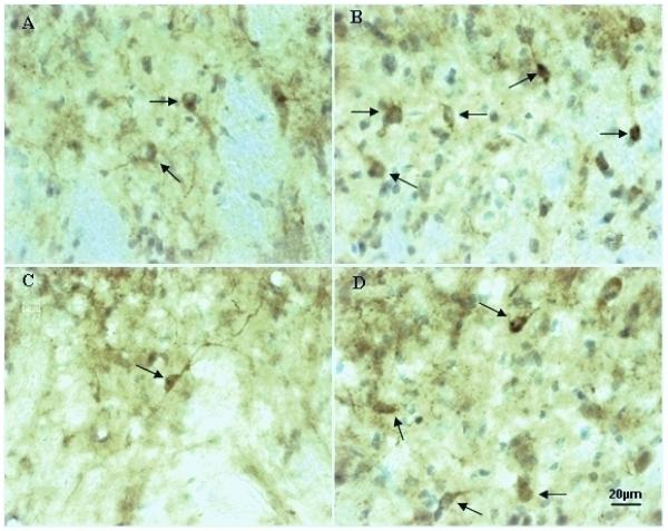
Fig. 5.
The high magnification micrographs show nNOS immunoreactivity in the gracile nucleus of a LC rat (A) and a ZDF rat (C) compared to a LC rat (B) and a ZDF rat (D) with 2,5-HD intoxication. nNOS immunostained cells (indicated by arrows) were increased in the ipsilateral side of the gracile nucleus in a LC rat and a ZDF rat (B and D) with 2,5-HD treatment (B) compared to a LC rat and a ZDF rat without treatment (A and C). nNOS immunoreactivity was also reduced in the gracile nucleus of a control ZDF rat (C) compared to a LC rat (A).

