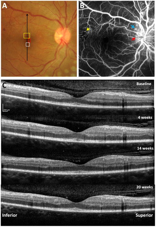
Figure 2.
Clinical retinal images of a 35 year old male PDR patient with T2DM. A) Fundus photograph with regions of interest on the right eye imaged by AOSLO, as presented in Figures 3, 4 and 10. Black arrow indicates location of vertical SD-OCT scanning. B) Intravenous fluorescein angiography showing scattered MAs with a temporal region of capillary nonperfusion (yellow arrow) and neovascularisation (red arrow) with leakage (blue arrow) at and around the disc. C) Vertical SDOCT scans through the fovea across the four visits.

