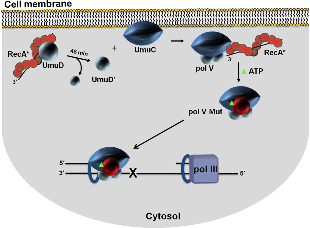
Figure 7.
Spatial regulation of pol V Mut activity in vivo. Sketch depicting cell membrane localization of UmuC. Single molecule live cell fluorescence imaging data indicate that after an initial delay post UV irradiation, UmuC is synthesized and localized on the inner cell membrane. UmuC is released into the cytosol only after it interacts with UmuD'2. As RecA and UmuD are found in or near the membrane, additional steps in the formation of pol V Mut (UmuD'2C-RecA-ATP) may occur in proximity to the membrane. It is not clear what functionality may be associated with the RecA protein found in the membrane. Active RecA*, as studied to date, consists of RecA protein filaments bound to DNA. Sequestration of UmuC at the membrane provides additional levels of pol V Mut regulation. A delay between the production of UmuC and the formation of cytosolic pol V Mut provides time for error-free DNA repair to occur before mutagenic pol V Mut lesion bypass is necessary.

