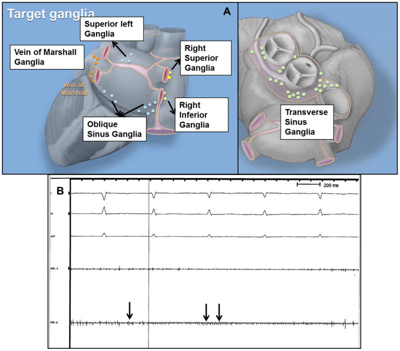
Figure 1.

(A) Location of major ganglia targeted for ablation. (B) Ganglion potentials are seen over the transverse sinus ganglionated plexus in sinus rhythm. Ganglion potentials are characterized by high frequency signal (arrows) that are dissociated from the cardiac cycle and occur stochastically. Atrial electrograms are not seen prominently in this tracing. ABL, d and p – distal and proximal bipoles of quadripolar catheter on fat pad.
