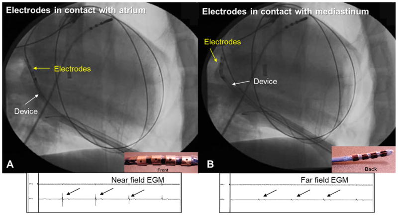
Figure 3.

Electrogram guided ablation. The quadripolar catheter is positioned in the oblique sinus. (A) When the electrodes are in contact with the atrium, high amplitude near field atrial electrograms (EGM) is noted and ablation was safely performed. (B) When the electrodes are in contact with the pericardium in the oblique sinus, atrial electrograms are low amplitude and far field. The catheter was repositioned to avoid injury to the esophagus.
