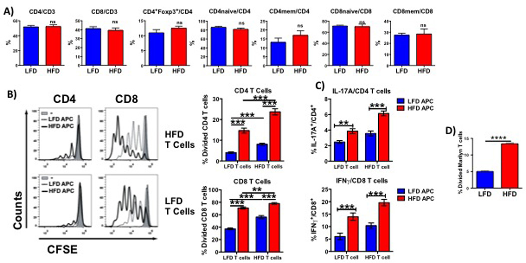
Figure 2. HFD results in greater T cell activation.

Percentages of CD4+, CD8+, Tregs, memory and naïve T cells (A) in splenocytes from HFD and LFD mice were analyzed by flow cytometry. Proliferation (B), IFNγ production by CD8+ cells (C) and IL-17 expression by CD4+ cells were assessed in HFD and LFD T cells stimulated with HFD or LFD APCs. (D) Proliferation of CFSE-labeled CD4+ Marilyn T cells stimulated for 3 days with APCs isolated from mice maintained on HFD or LFD for >12 weeks. Experiments were performed in groups of at least 3 mice and repeated at least twice. Results are displayed as mean +/− SEM. Comparisons were performed using a two-tailed unpaired t test. **: p<0.01, ***: p<0.001, ****: p<0.0001, ns: not significant.
