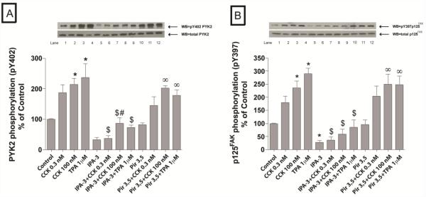
Fig. 2. Effect of IPA-3 and Pir 3,5 on the ability of physiological (0.3 nM) and supraphysiological (100 nM) concentrations of CCK and TPA (1 μM) to stimulate various kinases of the focal adhesion pathway (PYK2 and p125FAK).
Rat pancreatic acinar cells were pretreated with no additions or with IPA-3 (40 μM) or Pir 3,5 (40 μM) for 15 min and then incubated with no additions (control), with 0.3 nM CCK or 100 nM CCK for 3 min or with 1 μM TPA for 5 min and then lysed. Whole cell lysates were submitted to SDS-PAGE and transferred to nitrocellulose membranes. Membranes were analyzed using anti-pY402 PYK2 and anti-pY397 p125FAK. Antibodies detecting total amount of these kinases were used to verify loading of equal amounts of protein. The bands were visualized using chemoluminescence and quantification of phosphorylation was assessed using scanning densitometry. Both a representative experiment of 4 others and the means of all the experiments are shown. * P< 0.05 vs. control, # P< 0.05 vs. IPA-3 alone, ∞ P< 0.05 vs. Pir 3,5 alone and $ P< 0.05 comparing stimulants (CCK or TPA) preincubated with 1% DMSO vs. stimulants pre-incubated with IPA-3 or Pir 3,5, respectively.

