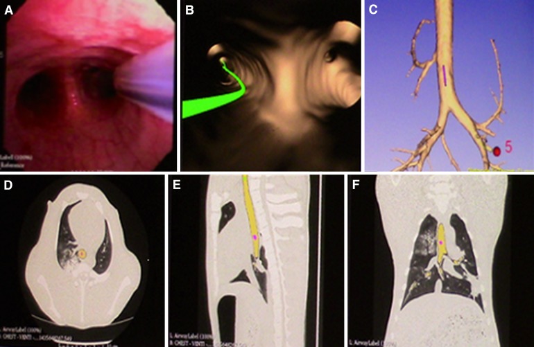
Fig. 3.
Our electromagnetic navigation system multiplanar window. a Real-time bronchoscopy image and the sensor probe. b Virtual bronchoscopy image and the navigation pathway (green line) toward the target lesion. c 3D tracheobronchial anatomical model. d, e, f Axial, sagittal and coronal images displaying the sensor location in the computerized tomography images [25]

