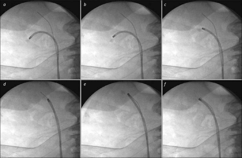
Figure 3.
Basket relocation of lower pole stone. 3a. The stone has been identified in the lower pole, and is grasped in a basket to prepare for relocation. 3b and c. The ureterorenoscope is manoeuvred out of the lower calyx towards the renal pelvis. 3d. The scope is advanced into the patient to the upper pole – the presence of the safety wire in the upper calyx can aid this both under endoscopic and fluoroscopic control. 3e. The stone is then released from the basket, which is withdrawn from the scope, and replaced with an appropriate laser fibre for stone fragmentation. 3f. The stone has been successfully broken into small pieces. It will be easier to re-pass the ureterorenoscope back and forth to this upper calyx for basket stone retrieval than to direct it repeatedly to the lower calyx where the stone was originally, particularly as the safety wire “guides the way”.

