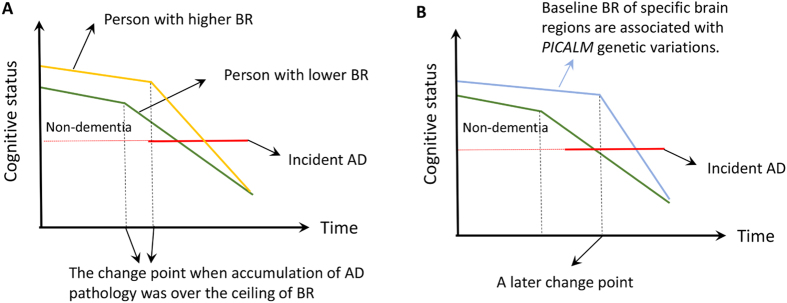
Figure 1.
(A) Depiction of how brain reserve operates to protect the brain. The x-axis represents time, over which AD pathology slowly accumulates. The y-axis represents cognitive function. We assume that AD pathology accumulates over time at the same rate in two individuals with high and low brain reserve (BR). The amount of pathology needed before cognitive function is affected is greater for individual with higher CR, leading to a later change point of time. It follows that greater pathology will be needed for the person with higher BR to meet clinical diagnostic criteria for AD, thus delaying the onset of the disease. Once cognitive decline arises, it is faster in the person with higher BR22. (B) We proposed a hypothesis that PICALM genetic variations were associated with brain reserve (baseline thickness/volume and atrophy rate) of specific regions associated with AD in non-dementia elderly. We hypothesized that individuals carrying specific PICALM variations might have higher baseline thickness/volume of specific brain areas and/or slower atrophy rate in confrontation with impacts of pathological impairments and/or normal aging for some unknown reasons. Based on model depicted in (A), these trends equivalently render two powerful “weapons” for the individuals to maintain normal cognition and stay away from AD over a period longer than others. Abbreviation: BR = brain reserve; AD = Alzheimer’s disease.

