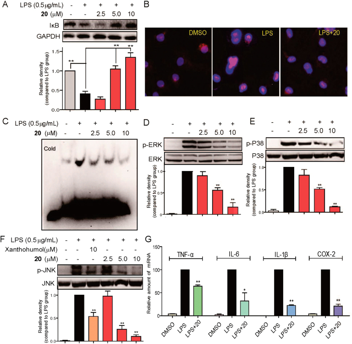
Figure 4. Effects of compound 20 on the LPS-induced activation of MAPK and NF-κB and expression of inflammatory genes.
(A,D,E,F) MPMs pretreated with vehicle or compound 20 (2.5, 5, or 10 μM) or Xanthohumol (10 μM) for 30 min followed by incubation with LPS (0.5 μg/mL) for 20 min. The protein levels of IκB, GAPDH, p-ERK, ERK, p-P38, P38, p-JNK and JNK were measured by immunoblot analysis. (B) MPMs pretreated with vehicle or compound 20 (10 μM) for 30 min followed by incubation with LPS (0.5 μg/mL) for 1 h. Cells were subjected to fluorescence microscopy (200×), displaying the NF-κB P65-stained with Cy3-labeled antibody in pink and the nuclei-stained with 4,6-diamidino-2-phenylindole in blue. (C) MPMs pretreated with vehicle or compound 20 (2.5, 5, or 10 μM) for 30 min followed by incubation with LPS (0.5 μg/mL) for 1 h. Nuclear extracts were analyzed for NF-κB activity by EMSA. (G) MPMs pretreated with vehicle or compound 20 (10 μM) for 30 min followed by incubation with LPS (0.5 μg/mL) for 6 h. Expressions of TNF-α, IL-6, IL-1β, and COX-2 were analyzed with quantitative PCR (qPCR) using specific primers and normalization against the housekeeping gene β-actin. Data are mean values (±SEM) of 3–5 separate experiments. *p < 0.05 and **p < 0.01 vs. only-LPS stimulated group. The gels were run under the same experimental conditions. Shown are cropped gels/blots (The gels/blots of 4A and 4D-F with indicated cropping lines are shown in Supplementary Figure S8).

